|
|
Cervical Aortic Arch
General Considerations
- Rare congenital anomaly
- Usually defined as a supraclavicular position of the aortic arch
- More common on the right side
- 2:1 female:male predominance
Clinical Findings
- Pulsatile neck mass
- Stridor
- Dyspnea
- Dysphagia
- Respiratory infections
Haughton Classification of Cervical Aortic Arches |
Type A |
Contralateral descending aorta and absence of one common carotid artery (separate external and internal carotid artery branches) |
Type B |
Contralateral descending aorta and presence of both common carotid arteries |
Type C |
Contralateral descending aorta and bi-carotid trunk |
Type D |
Ipsilateral descending aorta with normal sequence of brachiocephalic branching |
Type E |
Right aortic arch and right descending aorta |
Imaging Findings
- Apical mass
- Absence of the normal aortic knob
- Displacement of the trachea to the side opposite the arch
- Right-side cervical arches usually descend on the left
- Anomalous origins of the great vessels
- Right-sided lesions
- Right apical mass-like density
- Absence of aortic knob on left
- Aorta usually descends on left (80%)
- Displace the trachea and esophagus forward
- Branching of major vessels may be mirror-image
- Left-sided lesions
- Aortic knob appears at apex of left lung
- Aorta usually descends on the left (70%)
- Do not usually displace the trachea and esophagus forward
Differential Diagnosis
- Buckling of the aorta from a pseudocoarctation may present a similar picture
- Aneurysms of the great vessels
Associations
- Right-sided lesions have been associated with the 22q11.2 deletion syndrome
- Right-sided lesions more often associated with intracardiac abnormalities like VSD or conotruncal abnormalities
- Left-side lesions more often associated with coarctation
Treatment
- Surgical treatment may be indicated for relief of tracheal and esophageal compression symptoms or for correction of associated vascular defects


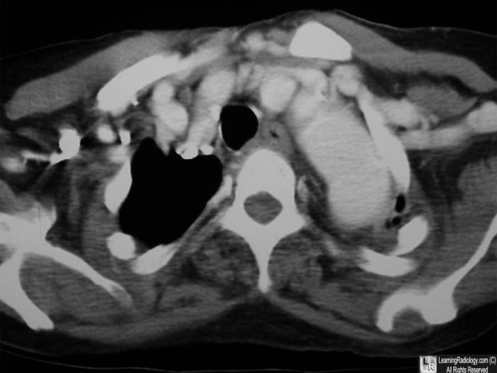
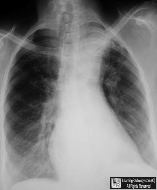
Cervical Aortic Arch. Above: Axial, contrast-enhanced CT scan of the upper thorax shows the aortic arch (black arrow) extending above the level of the clavicles. Below: Frontal radiograph of the chest demonstrates the cervical arch at the apex of the left lung (white arrow) above the clavicles. There is no aortic arch in the normal location. The aorta descends on the left.
For this same photo without the arrows, click here and here
For more information, click on the link if you see this icon 
The cervical aortic arches. VM Haughton, KE Fellows and AE Rosenbaum. Radiology. 1975 Mar;114(3):675-81.
The Cervical Aortic Arch. R Moncada, M Shannon, R Miller, H White, J Friedman, And Wh Shuford. Ajr November 1975 Vol. 125no. 3 591-601
Human Malformations And Related Anomalies. RE Stevenson and JG Hall. Oxford University Press, 2006
|
|
|
